
First IR and X-ray micro-tomography of asteroidal grains
How to measure and visualize in 3D the composition and structure of a micrometric extraterrestrial grain without destroying it? Researchers from the "Astrochemistry and Origin" team of the IAS and from the SOLEIL synchrotron, in collaboration with two Japanese laboratories, have shown that this is possible. Coupling IR and X-ray micro-tomography the scientist analyzed samples from the asteroid Itokawa, collected by the Hayabusa probe (JAXA).
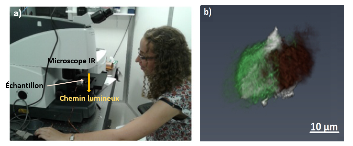
For many years the "Astrochemistry and Origins" team of the IAS has been specializing in the study of small bodies of the solar system (asteroids, comets and interplanetary grains). In particular, the team has developed a strong expertise on laboratory infrared (IR) spectroscopy of extraterrestrial grains. Lately, thanks to technological advances in IR detectors, the team has been able to apply IR hyper-spectral spectroscopy in 3D at the micrometric scale.
X-rays micro-tomography is a 3D imaging technique that provides information on the sample’s physical properties (such as the shapes of the various constituents, its porosity and 3D assembly). While X-ray micro-tomography is a well-established technique in many research fields (and particularly in medicine), infrared micro-tomography is a new method. Recently installed at the SOLEIL synchrotron, thanks to an international collaboration led by the Astrochemistry and Origin team of the IAS and the SMIS team of SOLEIL, infrared micro-tomography allows to probe the 3D composition of micrometric samples in a completely non-destructive way. This technique is complementary to X-ray micro-tomography, as it is sensitive to the molecular vibrations and provides information on the 3D distribution of organics and minerals and their local degree of hydration.
For the first time, researchers from the "Astrochemistry and Origin" team have coupled IR and X-ray micro-tomographies, to analyze six extraterrestrial grains. The IR analysis was conducted at SMIS-SOLEIL (France), the X-ray analyses were performed at the PSICHE beamline of SOLEIL and at the beamline BL47XU of the SPring-8 synchrotron (Japan). These grains, with diameters between 30 and 50 µm, have two origins:
(i) Five grains were collected by the Hayabusa mission (JAXA), directly on the surface of the asteroid Itokawa.
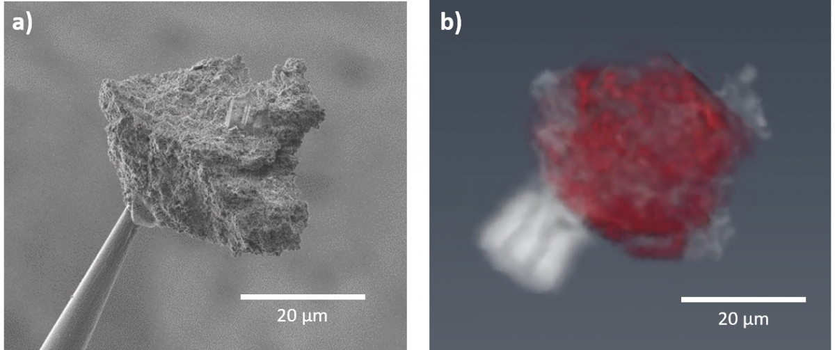
(ii) A micrometric fragment of the Paris meteorite, one of the least altered meteorites on Earth. This sample was found to be particularly interesting because it contains aliphatic organic matter.
The analysis of the Itokawa samples proved it is possible to apply the combination of IR and X-ray micro-tomographies on grains retrieved by sample return missions (Fig. 1). Combining these techniques allowed to study the mineral composition and the 3D assembly of the asteroid samples.
The study of the organic-rich Paris meteorite proved that the heterogeneous 3D spatial distribution of aliphatic organic matter and its composition can be detected through non-destructive techniques, and thus without altering the grain chemically or physically (Fig. 2).
The latest sample return missions Hayabusa2 (JAXA) and OSIRIS-REx (NASA) target carbonaceous asteroids Ryugu and Bennu, respectively, and will be retrieving only a limited amount of material (a few grams at most). There is therefore the need to envisage an analytical strategy that maximizes the scientific outcomes while minimizing sample loss. The combination of IR and X-ray micro-tomographies can constitute the first phase of a multi-analytical protocol, as it allows to shed light on the 3D distribution of the organic and mineral phases of extraterrestrial matter in a non-destructive way.
Related articles:
- Z. Dionnet, R. Brunetto, A. Aléon-Toppani, S. Rubino, D. Baklouti, F. Borondics, A-C. Buellet, Z. Djouadi, A. King, T. Nakamura, A. Rotundi, C. Sandt, D. Troadec and A. Tsuchiyama, 2020. Combining IR and X-ray micro-tomography data sets: Application to Itokawa particles and to Paris meteorite. Meteoritics and Planetary Science 55, 1645-1664.
- A. Aléon-Toppani, R. Brunetto, J. Aléon, Z. Dionnet, S. Rubino, D. Levy, D. Troadec, F. Borondics and F. Brisset, 2020. A Preparation Sequence for Multi-Analysis of Micrometer-Sized Extraterrestrial Samples. Lunar and Planetary Science Conference, LPI Contribution No. 2326.
- R. Brunetto and C. Lantz, 2019. Laboratory perspectives on sample returns from Hayabusa2 and OSIRIS-REx. Nature Astronomy 3, 290–292.